
Case Study
Lightfield Microscopy for Zebrafish Brain Imaging
Increasing the spatial and temporal resolution of light-field microscopy by a combined factor of 100.
The Client
Development for the Lotus program was supported by a Small Business Innovation Research (SBIR) grant from the National Institute of Mental Health (NIMH) of the National Institutes of Health (NIH), under Award Number R43MH109332.
The NIH funds transformative biomedical technologies to accelerate scientific research and clinical breakthroughs. This project was supported by the NIMH, whose mission includes advancing tools for neuroscience research—such as high-performance embedded systems for real-time data acquisition and experimental control.
This program was a collaboration between LeafLabs and the Synthetic Neurobiology Group at MIT.
The Challenge
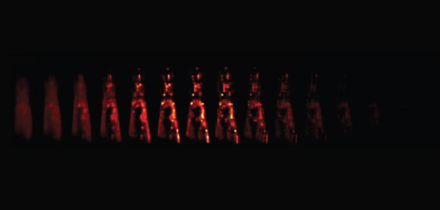
Light-field microscopy (LFM) is a functional, plenoptic imaging technique that offers ultrahigh spatial and temporal resolution for 3D tissue samples. Lotus’ goal was to use light-field microscopy to record spikes from every neuron in the zebrafish brain.
Capturing real-time imaging data at a staggering 125 Gbps from a custom optical system posed significant technical hurdles. The system needed to manage data rates equivalent to 250,000 channels of full-band electrophysiological data—approximately four times the data throughput of the ALICE detector at CERN. This required a robust architecture capable of synchronized acquisition from multiple high-speed sensors without data loss or latency.
Our Approach
The proposal called for the use of 16 custom FPGA boards (based on the Willow architecture) recording in sync from an array of CMOS sensors. Each board interfacing with sCMOS sensors, capturing and processing data in parallel to handle the throughput. Key components of our approach included:
Optical System Integration: Designing and aligning a custom optical pathway incorporating long working distance objectives, large tube lenses, and microlens arrays to optimize light-field capture.
High-Speed Data Acquisition: Implementing FPGA-based pipelines to manage real-time data streams from multiple sCMOS sensors, ensuring synchronized acquisition and minimal latency.
Data Management: Developing firmware and software solutions for efficient data handling, storage, and preliminary processing to facilitate downstream analysis.
User Interface Development: Creating intuitive interfaces for system control, monitoring, and data visualization to support experimental workflows.
Impact
Lotus significantly advances the capabilities of light-field microscopy, achieving a combined 100-fold increase in spatial and temporal resolution. This platform enables comprehensive, real-time imaging of neuronal activity, providing researchers with unprecedented insights into brain function. The successful integration of high-speed optics, data acquisition, and processing sets a new standard for functional imaging systems.
By overcoming the challenges associated with ultra-high-speed data capture and processing, Lotus empowers neuroscientists to observe and analyze complex neural dynamics with unparalleled detail. This capability is crucial for advancing our understanding of brain function and developing interventions for neurological disorders.
Tech Stack
sCMOS Image Sensors
Custom Firmware in C, Python
PCIe, Ethernet
FreeRTOS
Data Visualization and Analysis Tools
Capabilities
Custom Optical System Design
High-Speed Data Acquisition
FPGA Development and Integration
Firmware and Software Engineering
System Synchronization and Control
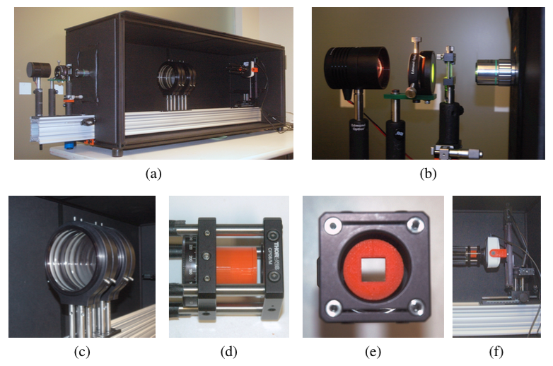
Experimental prototype showing the long working distance objective, large tube lens, microlens array and camera sensor.
Tackling challenges in real-time imaging or data acquisition?
Let's explore how LeafLabs’ expertise can drive your project forward.

Lotus Resources
Publication: Robert Prevedel, etal. "Simultaneous whole-animal 3D imaging of neuronal activity using light-field microscopy". Nature Methods, 22 April 2014
For more information about Lotus, please contact us at neuro@leaflabs.com